Advanced single-cell transcriptomic analysis of the lung tumor microenvironment using Scanpy, scVI-tools, LIANA, pathway enrichment analysis, and trajectory inference.
This project reconstructs cellular organization, cell–cell communication networks, epithelial differentiation programs, and metastatic adaptations using the GSE131907 lung tumor microenvironment dataset.
The tumor microenvironment consists of complex interactions between malignant, immune, and stromal cell populations that influence tumor progression, metastasis, and therapeutic response.
This project implements a complete modern single-cell RNA-seq workflow to characterize cellular heterogeneity and biological signaling within lung tumors.
The analysis includes:
- Single-cell quality control and preprocessing
- Batch integration using scVI
- Cell type and subtype annotation
- Differential expression analysis
- Pathway enrichment analysis
- Cell–cell communication inference
- Pseudotime trajectory reconstruction
- Metastatic versus normal tissue comparison
✔ Single-cell RNA-seq analysis
✔ scVI latent-space integration
✔ Cell-type annotation and subtype discovery
✔ Differential expression analysis
✔ GO and KEGG enrichment analysis
✔ LIANA cell–cell communication inference
✔ Diffusion pseudotime trajectory analysis
✔ Metastatic microenvironment characterization
✔ Reproducible Scanpy workflow
Major cell populations identified included:
- epithelial cells
- endothelial cells
- fibroblasts
- myeloid cells
- NK cells
- T lymphocytes
- B lymphocytes
- oligodendrocytes
- mast cells
Ligand–receptor analysis identified strong interactions including:
- ARPC5 → ADRB2
- ACTR2 → ADRB2
- APP → AGER
- APOE → ABCA1
These interactions suggest active communication between epithelial, immune, and stromal compartments.
Pseudotime analysis revealed:
- continuous epithelial differentiation programs
- terminal states enriched for AGER and AQP5
- AT1-like alveolar epithelial signatures
Brain metastatic samples showed enrichment of:
- APLP1
- APOD
- AIF1L
- ANLN
- ABCA2
Functional enrichment suggested adaptation through:
- vesicle trafficking
- phagosome maturation
- intracellular pH regulation
- metastatic microenvironment remodeling
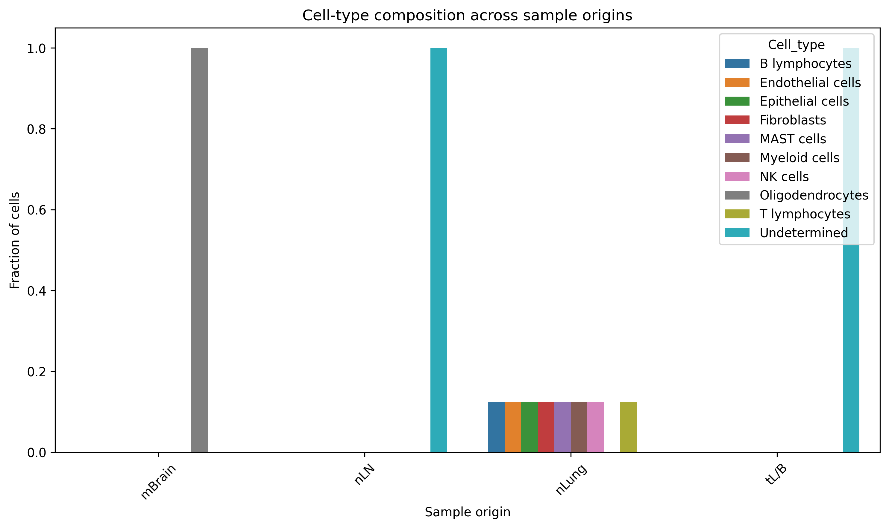
Cellular composition across normal lung, lymph node, tumor, and metastatic samples.
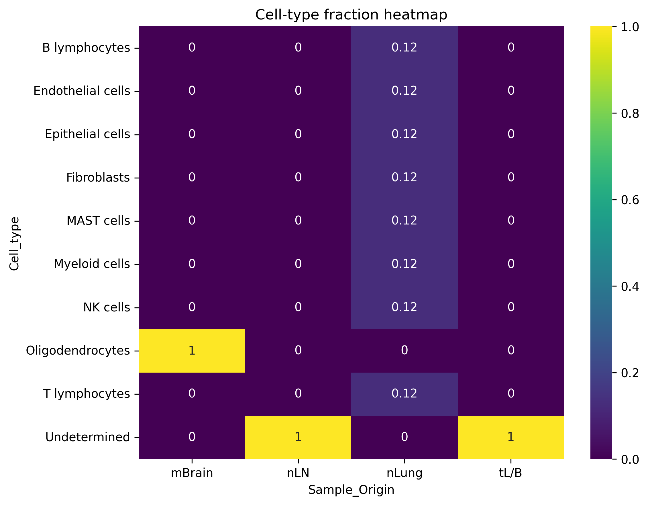
Relative abundance of cellular populations across biological compartments.
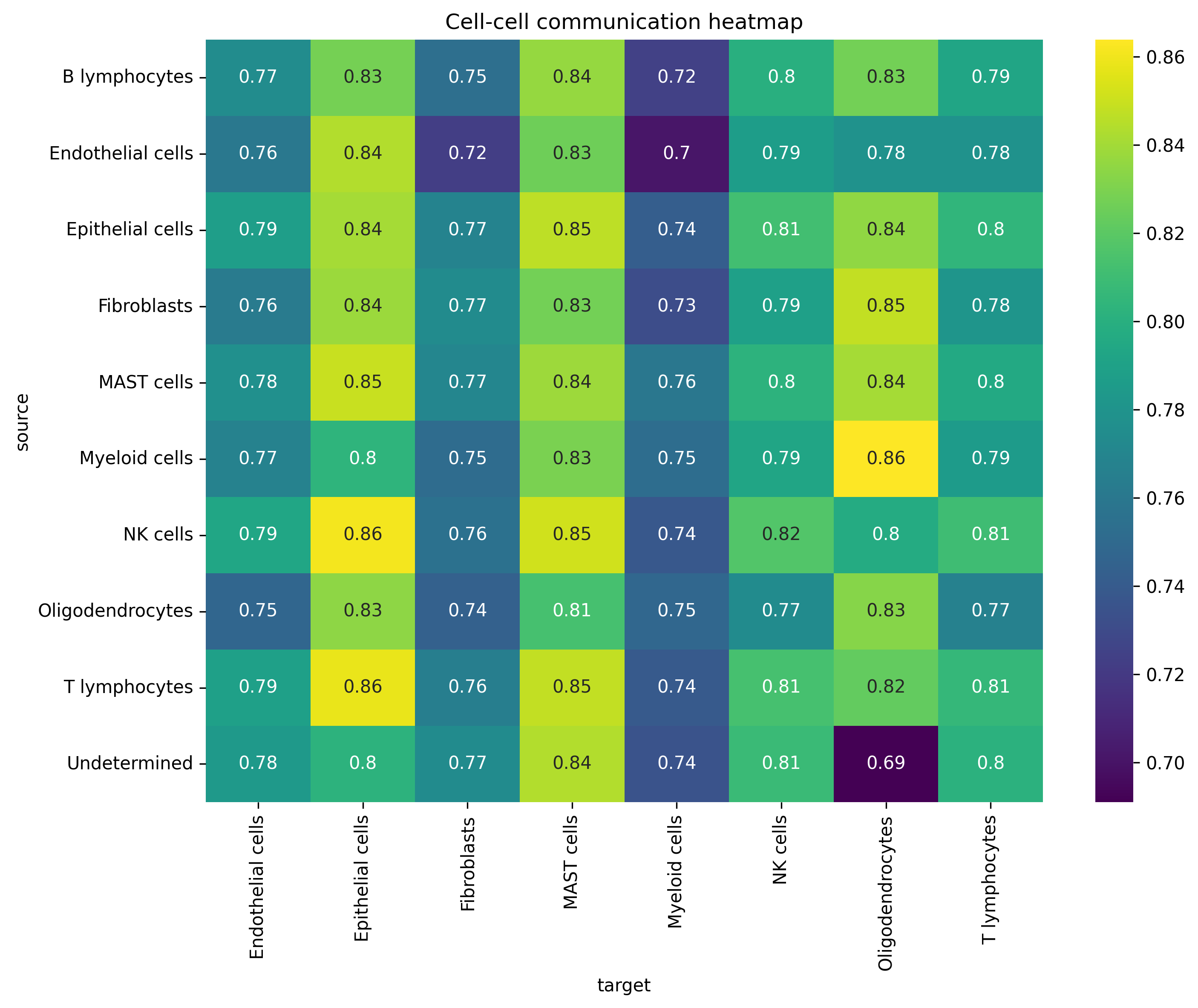
Global signaling interactions inferred using LIANA.
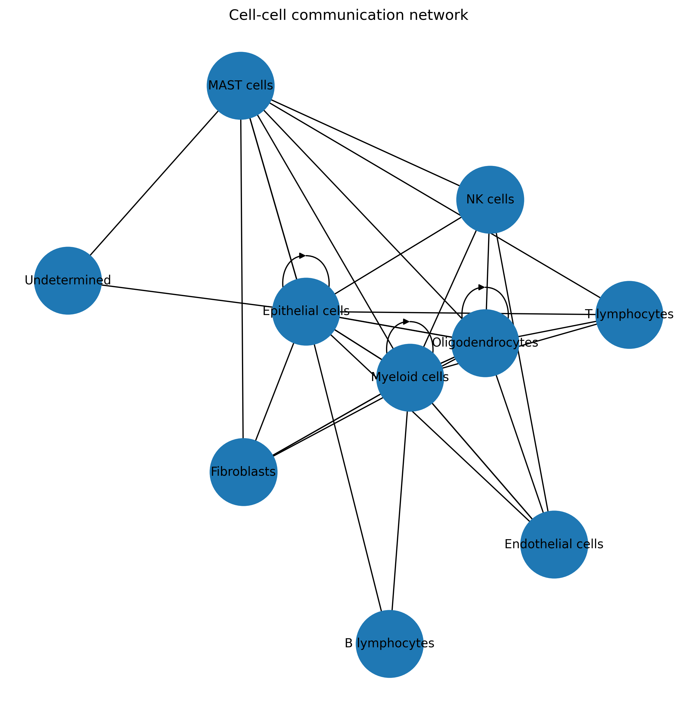
Network representation of intercellular communication.
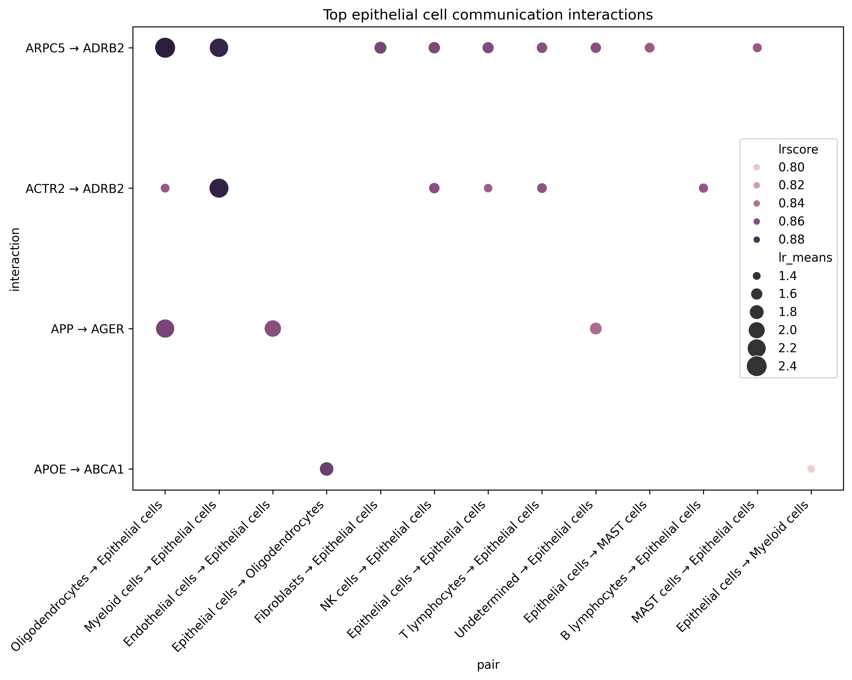
Top epithelial ligand–receptor interactions.
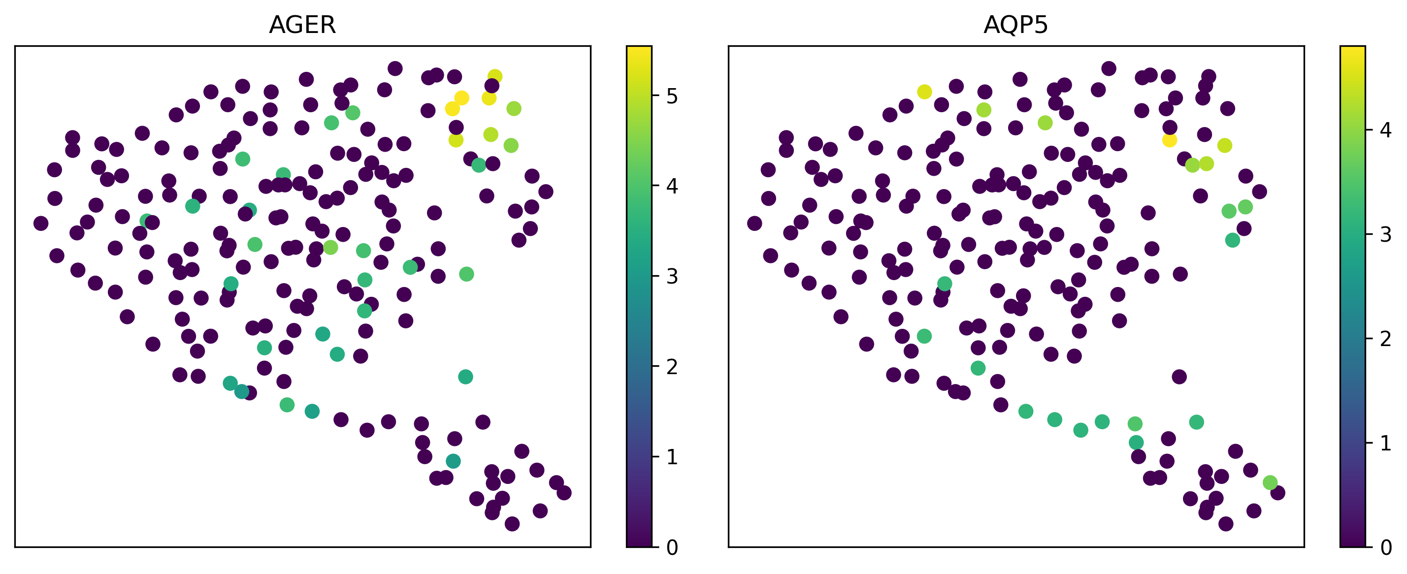
Gene-expression changes along epithelial differentiation trajectories.
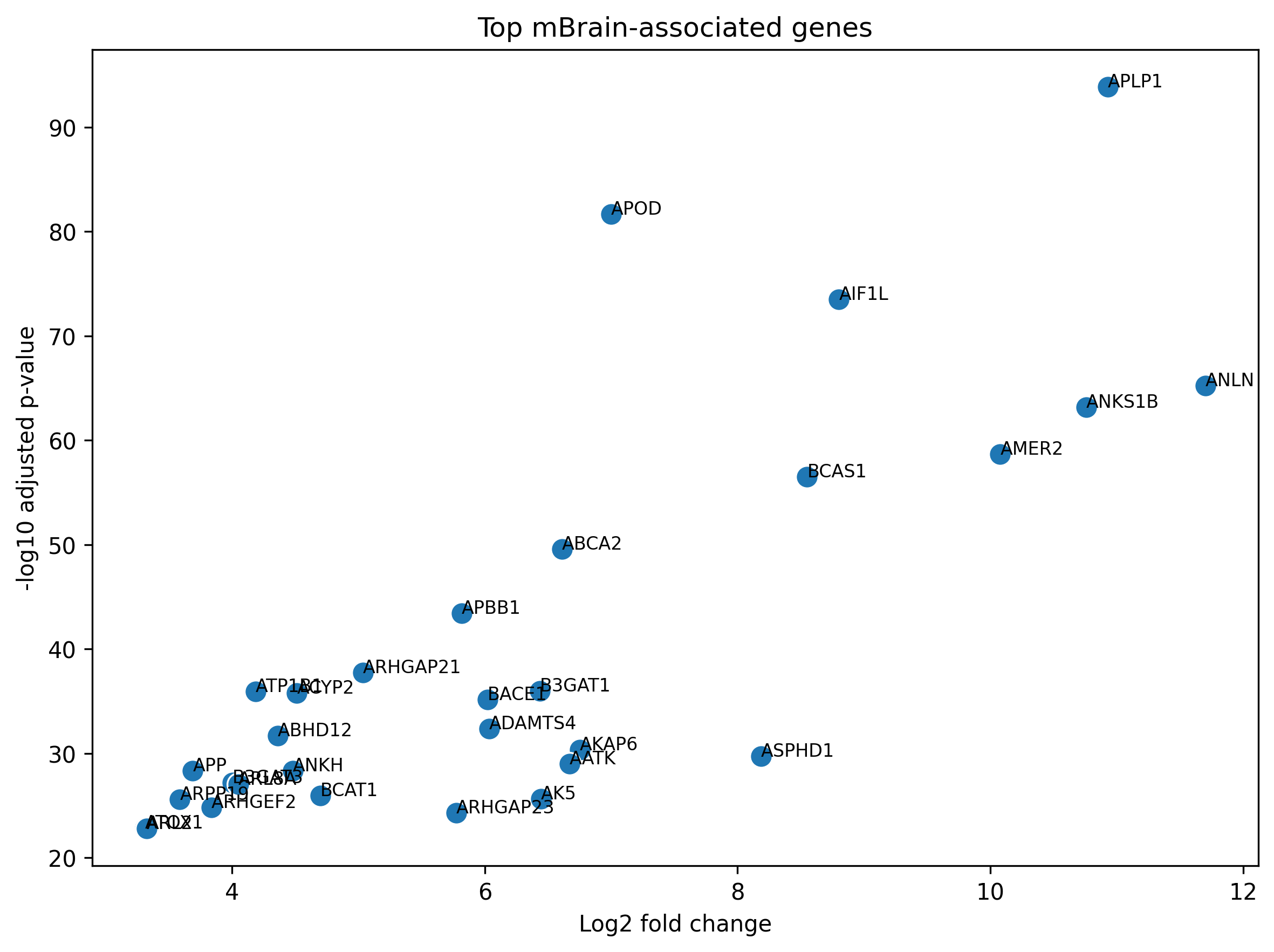
Genes enriched in brain metastatic samples.
GEO Accession: GSE131907
Sample origins:
- nLung — normal lung
- nLN — normal lymph node
- tL/B — tumor lung / bronchus
- mBrain — brain metastasis
- Low-quality cell filtering
- Mitochondrial QC
- Gene and cell thresholding
- Normalization
- Log transformation
- Highly variable gene selection
- PCA and neighborhood graph construction
- Batch correction
- Latent space learning
- Integrated UMAP generation
- Leiden clustering
- Cell-type annotation
- Subtype characterization
- Marker gene identification
- Wilcoxon testing
- Biological interpretation
- GO enrichment
- KEGG enrichment
- Functional annotation
- LIANA ligand–receptor analysis
- Communication networks
- Interaction heatmaps
- Diffusion pseudotime
- PAGA graph analysis
- Epithelial lineage reconstruction
- Sample-origin comparison
- Brain metastasis signatures
- Functional enrichment
multimodal-cancer-foundation-models/
│
├── data/
├── figures/
├── results/
├── notebooks/
│   ├── 01_quality_control.ipynb
│   ├── 02_preprocessing.ipynb
│   ├── 03_scvi_integration.ipynb
│   ├── 04_clustering_annotation.ipynb
│   ├── 05_differential_expression.ipynb
│   ├── 06_pathway_enrichment.ipynb
│   ├── 07_cell_communication.ipynb
│   ├── 08_trajectory_analysis.ipynb
│   └── 09_multisample_analysis.ipynb
│
└── README.md
- Scanpy
- AnnData
- scVI-tools
- LIANA
- GSEApy
- GO enrichment
- KEGG enrichment
- Python
- pandas
- NumPy
- matplotlib
- seaborn
- scRNA-seq preprocessing
- Batch integration
- Cell annotation
- Differential expression
- Tumor microenvironment analysis
- Metastatic biology
- Cell-state characterization
- Tumor heterogeneity
- Cell–cell communication
- Pathway enrichment
- Trajectory inference
- Biological interpretation
- High-dimensional data analysis
- Reproducible research
- Scientific visualization
- Large-scale transcriptomics
- Spatial transcriptomics integration
- RNA velocity analysis
- CNV inference
- NicheNet and CellChat comparison
- Survival-associated cell-state signatures
- Foundation model integration for multimodal biology
Agata Gabara
MSc Bioinformatics Student
Research Interests:
- Single-Cell Transcriptomics
- Cancer Genomics
- Tumor Microenvironment Biology
- Computational Oncology
- Multi-Omics Integration
GitHub: https://github.com/ag48665
LinkedIn: https://www.linkedin.com/in/agatha-gabara-06494a37/