MRI_Fibrosis_Tool
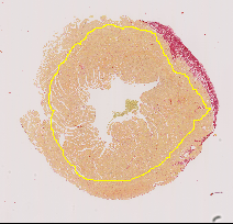
Measure the relative area of sirius red stained fibrosis. The tool uses the colour deconvolution plugin from Gabriel Landini.
You can find test images here and here.
To install the tool, drag the link MRI_Fibrosis_Tool.ijm to the ImageJ launcher window, save it under macros/toolsets in the ImageJ installation.
Select the "MRI_Fibrosis_Tool" toolset from the >> button of the ImageJ launcher.

- the first button (the one with the image) opens this help page.
- the m-button runs the analysis on the active window. Note that you should crop the image and that there should be a selection (roi) on the cropped image.
- the b-button runs the analysis on a folder containing cropped images with a selection (roi) in tif-format.
Right-click on the m-button to open the general options dialog:

- r1, g1, b1 - the RGB-components (between 0 and 1) of the colour we want to segment.
- r2, g2, b2 - the RGB-components (between 0 and 1) of the first background colour.
- r3, g3, b3 - the RGB-components (between 0 and 1) of the second background colour.
- file ext. - the file extension (including the dot) of the input files
Right-click on the b-button to open the batch-options dialog:
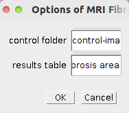
- control folder - the name of the sub-folder into which the control images will be saved
- results table - the name of the results table and the results spreadsheet file (csv text saved as .xls)
Colour deconvolution is used to separate the staining into a single image. A simple auto-threshold with the default method is applied and a selection is created. The area of the selection is measured and compared to the area of the selection on the input image. The colour deconvolution is done using the Colour Deconvolution Plugin1 from Gabriel Landini
Note that you can use the colour deconvolution plugin if you need to find different colour vectors. You can then enter the new values in the options dialog or change the default values in the macro's source code.
1: ImageJ.net - Colour Deconvolution


-
Tarasenko, N. et al. (2021) Valproic Acid Prodrug Affects Selective Markers, Augments Doxorubicin Anticancer Activity and Attenuates Its Toxicity in a Murine Model of Aggressive Breast Cancer, Pharmaceuticals, 14(12), p. 1244. doi:10.3390/ph14121244.
-
de Bakker, D.E.M., Bouwman, M., Dronkers, E., Simões, F.C., Riley, P.R., Goumans, M.-J., Smits, A.M., and Bakkers, J. (2021). Prrx1b restricts fibrosis and promotes Nrg1-dependent cardiomyocyte proliferation during zebrafish heart regeneration. Development 148, dev198937.
-
Hegemann, N., Primessnig, U., Bode, D., Wakula, P., Beindorff, N., Klopfleisch, R., Michalick, L., Grune, J., Hohendanner, F., Messroghli, D., et al. (2021). Right‐ventricular dysfunction in HFpEF is linked to altered cardiomyocyte Ca 2+ homeostasis and myofilament sensitivity. ESC Heart Failure 8, 3130–3144.
-
Osorio-Conles, Ó., Vega-Beyhart, A., Ibarzabal, A., Balibrea, J.M., Graupera, I., Rimola, J., Vidal, J., and de Hollanda, A. de (2021). A Distinctive NAFLD Signature in Adipose Tissue from Women with Severe Obesity. IJMS 22, 10541.
-
Kobayashi, H., Gieniec, K.A., Ng, J.Q., Goyne, J., Lannagan, T.R.M., Thomas, E.M., Radford, G., Wang, T., Suzuki, N., Ichinose, M., et al. (2021). Portal Vein Injection of Colorectal Cancer Organoids to Study the Liver Metastasis Stroma. JoVE 62630.
-
Dill, T.L., Carroll, A., Pinheiro, A., Gao, J., and Naya, F.J. (2021). The long noncoding RNA Meg3 regulates myoblast plasticity and muscle regeneration through epithelial-mesenchymal transition. Development 148, dev194027.
-
de Bakker, D.E.M., Dronkers, E., Bouwman, M., Vink, A., Goumans, M.-J., Smits, A.M., and Bakkers, J. (2020). Prrx1b directs pro-regenerative fibroblasts during zebrafish heart regeneration. bioRxiv.
-
Fischer, A., Bockstahler, M., Müller, A.-M., Stroikova, V., Leib, C., Pfitzer, G., Katus, H.A., and Kaya, Z. (2019). FN14 Signaling Plays a Pathogenic Role in a Mouse Model of Experimental Autoimmune Myocarditis. Journal of Cardiac Failure 25, 674–685.
-
Rabacal, W., Schweitzer, F., Rayens, E., Tarantelli, R., Whang, P., Jimenez, V.C., Outwater, J.A., and Norris, K.A. (2019). Statin treatment prevents the development of pulmonary arterial hypertension in a nonhuman primate model of HIV-associated PAH. Sci Rep 9, 19832.
-
Arunsan, P., Ittiprasert, W., Smout, M.J., Cochran, C.J., Mann, V.H., Chaiyadet, S., Karinshak, S.E., Sripa, B., Young, N.D., Sotillo, J., et al. (2019). Programmed knockout mutation of liver fluke granulin attenuates virulence of infection-induced hepatobiliary morbidity. ELife 8.
-
Romano, G., Reggi, S., Kutryb-Zajac, B., Facoetti, A., Chisci, E., Pettinato, M., Giuffrè, M.R., Vecchio, F., Leoni, S., De Giorgi, M., et al. (2018). APOA-1Milano muteins, orally delivered via genetically modified rice, show anti-atherogenic and anti-inflammatory properties in vitro and in Apoe atherosclerotic mice. International Journal of Cardiology 271, 233–239.
-
Rinrada Kietadisorn (2018). Drainage versus defense: The management of vascular leakage in cardiovascular diseases, Maastricht University.
-
Hollenbach, M., Thonig, A., Pohl, S., Ripoll, C., Michel, M., and Zipprich, A. (2017). Expression of glyoxalase-I is reduced in cirrhotic livers: A possible mechanism in the development of cirrhosis. PLOS ONE 12, e0171260.
-
MARCIO APARECIDO PEREIRA (2016). Tratamento com células derivadas do fígado embrionário retarda a progressão da fibrose hepática em ratos. Universidade de São Paulo.
 Volker Bäcker